A nanoscale mission represents a large leap ahead in understanding the human mind.
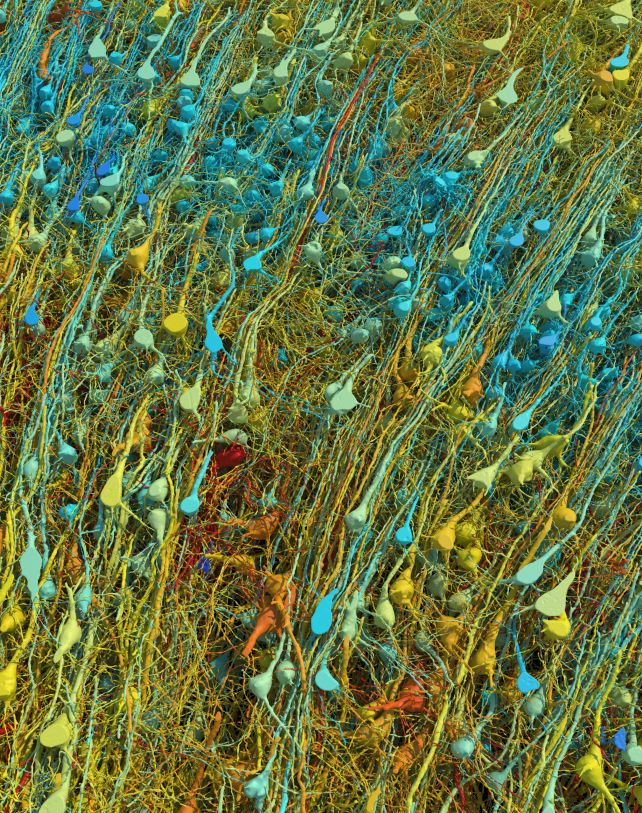
With greater than 1.4 petabytes of electron microscopy imaging information, a workforce of scientists has reconstructed a teeny-tiny cubic phase of the human mind.
It is only a millimeter on all sides – however 57,000 cells, 150 million synapses, and 230 millimeters of ultrafine veins are all packed into that microscopic area.
The work of virtually a decade, it is the most important and most detailed replica of the human mind so far all the way down to the decision of the synapses, the constructions that permit neurons to transmit alerts between them.
“The word ‘fragment’ is ironic,” says neuroscientist Jeff Lichtman of Harvard College. “A terabyte is, for most people, gigantic, yet a fragment of a human brain – just a miniscule, teeny-weeny little bit of human brain – is still thousands of terabytes.”
The human mind is notoriously complicated. Throughout the animal kingdom, the capabilities carried out by many of the very important organs are kind of the identical, however the human mind is in a league of its personal.
It is also very troublesome to check; there’s a lot occurring in there, on such miniscule scales, that we have been unable to know the synaptic circuitry intimately.
Every human mind comprises billions of neurons, firing alerts forwards and backwards by way of trillions of synapses, the command heart from which the human physique is run.
A deeper understanding of the way in which this dazzlingly difficult organ operates would confer profound advantages to our research of mind perform and problems, from harm to psychological sickness to dementia.
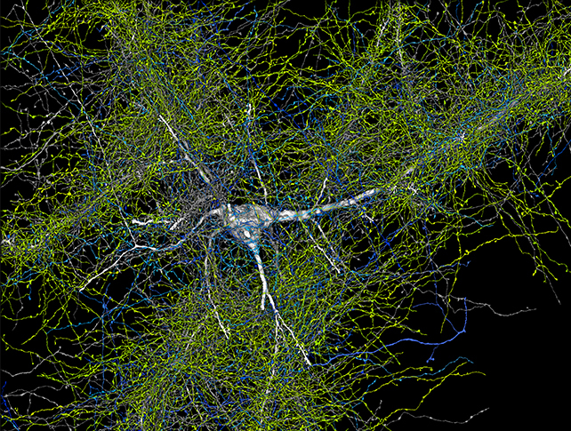
To that finish, Lichtman and colleagues have been engaged on what they name a “connectome” – a map of the mind and all its wiring that might assist higher perceive when that wiring is askew.
The present objective for the connectomics mission is the replica of a complete mouse mind, however utilizing related strategies to reconstruct at the very least segments of the human mind can solely advance our information sooner.
The workforce’s reconstruction was based mostly on a pattern of human mind excised from an epilepsy affected person throughout surgical procedure to entry an underlying lesion. The pattern was fastened, stained with heavy metals to intensify the small print, embedded in resin, and sectioned into 5,019 slices, with a imply thickness of 33.9 nanometers, collected on tape.
The researchers used high-throughput serial part electron microscopy to picture this tiny piece of tissue in mind-numbing element, producing 1.4 petabytes (1,400 terabytes) of information.
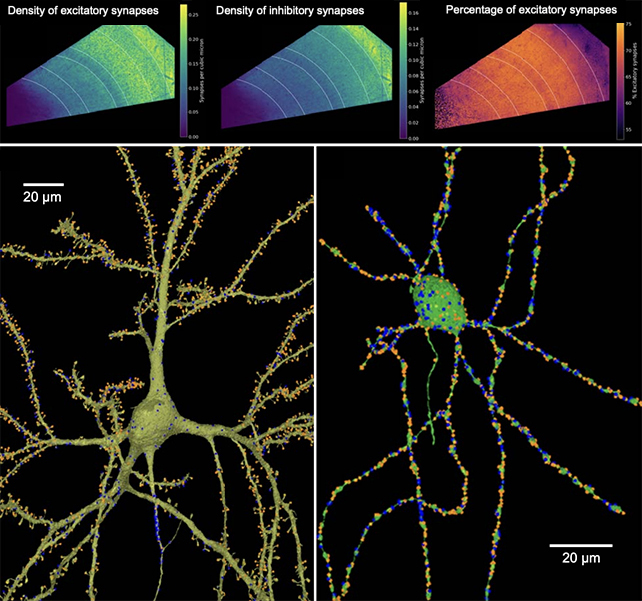
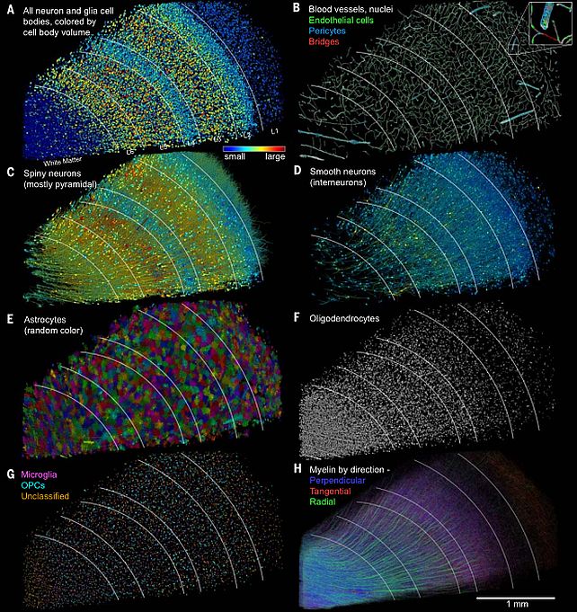
This information was analyzed with specifically developed strategies and algorithms, producing, the researchers say, “a 3D reconstruction of nearly every cell and process in the aligned volume.”
This reconstruction, named H01, has already revealed some beforehand unseen effective particulars in regards to the human mind. The workforce was shocked to notice that glia, or non-neuronal cells, outnumbered neurons 2:1 within the pattern, and the most typical cell kind was oligodendrocytes – cells that assist coat axons in protecting myelin.
Every neuron had hundreds of comparatively weak connections, however the researchers discovered uncommon, highly effective units of axons linked by 50 synapses. And so they discovered {that a} small variety of axons are organized in uncommon, intensive whorls.
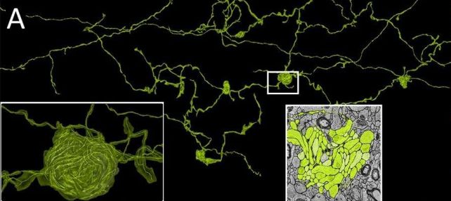
As a result of the pattern was taken from a affected person with epilepsy, it is unclear whether or not these are regular, however uncommon, options of the human mind, or linked to the affected person’s dysfunction. Both method, although, the work has revealed the huge breadth and depth of the chasm of our understanding of the mind.
The subsequent step within the workforce’s work entails making an attempt to know the formation of the mouse hippocampus, a mind area closely concerned in studying and reminiscence.
“If we get to a point where doing a whole mouse brain becomes routine, you could think about doing it in say, animal models of autism,” Lichtman defined final 12 months to The Harvard Gazette.
“There is this level of understanding about brains that presently doesn’t exist. We know about the outward manifestations of behavior. We know about some of the molecules that are perturbed. But in between the wiring diagrams, until now, there was no way to see them. Now, there is a way.”
The analysis has been printed in Science, and the information and reconstruction of H01 have been made freely accessible on a devoted web site.


